|  | 
IMAGING through turbid media has, in recent years, become a field of immense research, due to its great potential for diverse applications like imaging objects through fog, sighting aircraft through clouds, or searching for objects in murky waters. Aviation, defense, astrophysics, marine science are some of the areas which would benefit from advances in imaging through turbid media. Currently emphasis has mostly been on medical applications using infrared or visible light instead of harmful X-rays for imaging through tissues. Clinical studies have shown that the absorption characteristics at specific wavelengths by normal tissues differ significantly from some tumors and this property has been adapted for discriminating or locating tumors in the human body.
Light propagation in turbid media
Fog, milk and other random inhomogeneous media render imaging difficult due to the random multiple scattering of light. Inhomogeneities in the media cause scattering which may alter the direction of propagation, polarization and phase of light. Photons travel in straight line paths until they encounter an inhomogeneity, when they are scattered in random directions. The scattering is said to be in the Rayleigh regime when the radius, r, of the scatterer, is less than λ , the wavelength of light. In this case, the intensity of scattered light is equal in both the forward and backward directions (Figure 1 a). As the size of the particle increases, the scattering is more peaked in the forward direction (Figure 1 b). The case of r > λ , known as the Mie regime, shows peaking in some angles (Figure 1 c).
In a turbid medium made up of a random aggregate of scatterers, the photons undergo repeated scattering. The turbid medium is characterized by the scattering mean free path, ls, which is the mean distance the photons travel before getting scattered, and the transport mean free path, l*, which is the mean distance photons travel before the direction of propagation is randomized. Since it is quite possible that photons are forward scattered (and continue to travel in the same direction), l* > ls. The transport mean free path depends upon the number density of scatterers, refractive index contrast between the medium and the scatterers, and the anisotropy factor, i.e. a factor quantifying the directional distribution of scattering. Typical values of l* for infrared light in tissues are 1–2 mm.
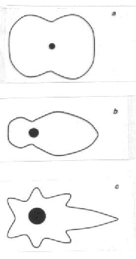 Figure 1. Intensity distribution of scattered light of wavelength λ from a spherical particle of radius a. r << λ ;b r = λ /4;c, r > λ . Figure 1. Intensity distribution of scattered light of wavelength λ from a spherical particle of radius a. r << λ ;b r = λ /4;c, r > λ .
The light emerging from a turbid medium consists of three components – the ballistic, the diffusive and the snake photons. These differ in their paths through the medium, and consequently in their imaging properties. The unscattered or forward scattered photons travel undeviated and emerge the first, having traveled the shortest distance through the medium (Figure 2). These preserve the characteristics of the incident light, namely direction of propagation, polarization and are hence best suited for imaging.
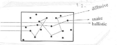 Figure 2. Trajectories of photons in a random medium, showing the ballistic, diffusive and snake components. Figure 2. Trajectories of photons in a random medium, showing the ballistic, diffusive and snake components.
However, they are few in number, their intensity falling off as I = Ioexp(–x/l*s) where Io is the input intensity, and x is the distance traveled. The diffuse component, which forms the bulk of the emergent light in turbid media is made up of photons that have undergone random multiple scattering, and these emerge later than the ballistic photons because of their increased path lengths. Their polarization, direction of propagation and phase are completely randomized. For most techniques of imaging, these form the unwanted, incoherent, diffuse background. Snake photons are those that travel in near-forward paths, having undergone few scattering events, all of which are in the forward or near-forward direction. Consequently, they retain the image bearing characteristics to some extent. Due to the varying path lengths the different photons have in the medium, a pulse of light incident on a turbid medium emerges elongated. Typically, a femtosecond pulse is stretched to a picosecond or a nanosecond pulse on traveling through a turbid medium.
All methods of imaging attempt to separate the undeviated ballistic or snake photons from the multiply scattered diffused photons; however, they differ in the method used to achieve this discrimination. While most methods use the ballistic photons to form shadowgrams of objects hidden in turbid media, a few others make use of the diffuse photons to infer about the objects. The rapid advance in this field over the last few years has been possible due to the technological advances, like the availability of femtosecond lasers, fast time-gated cameras and electronics.
Principles of imaging
The ballistic photons, having travelled a straight, undeviated path in the medium, can form shadows of objects in their path. Thus, if the ballistic photons could be separated from the diffusive, one could obtain shadowgrams of hidden objects. While there are several means of discriminating the ballistic photons in the presence of the diffusive, the most obvious technique appears to be time-gating, which exploits the fact that the diffusive photons arrive later than the ballistic photons. A short, femtosecond pulse is incident on the sample, and the emergent pulse sliced; the first femtosecond slice would form the shadowgram of the object hidden in the turbid medium.
PUBLICATIONS:
Time-gated backscattered ballistic light imaging of objects in turbid water Manuel E. Zevallos, S. K. Gayen, M. Alrubaiee, and R. R. Alfano APPLIED PHYSICS LETTERS 86, 1 (2005)
|  |  |